A collaborative research project between Chemistry, Radiology, Dermatology, Surgical Oncology, and Mathematics (NIH R01-CA166555) revealed that it is possible to detect nicotine in melanin in existing melanoma samples. This was an interesting development because literature suggests that smokers have a decreased risk of melanoma. However, this fell outside of the original scope of the research.
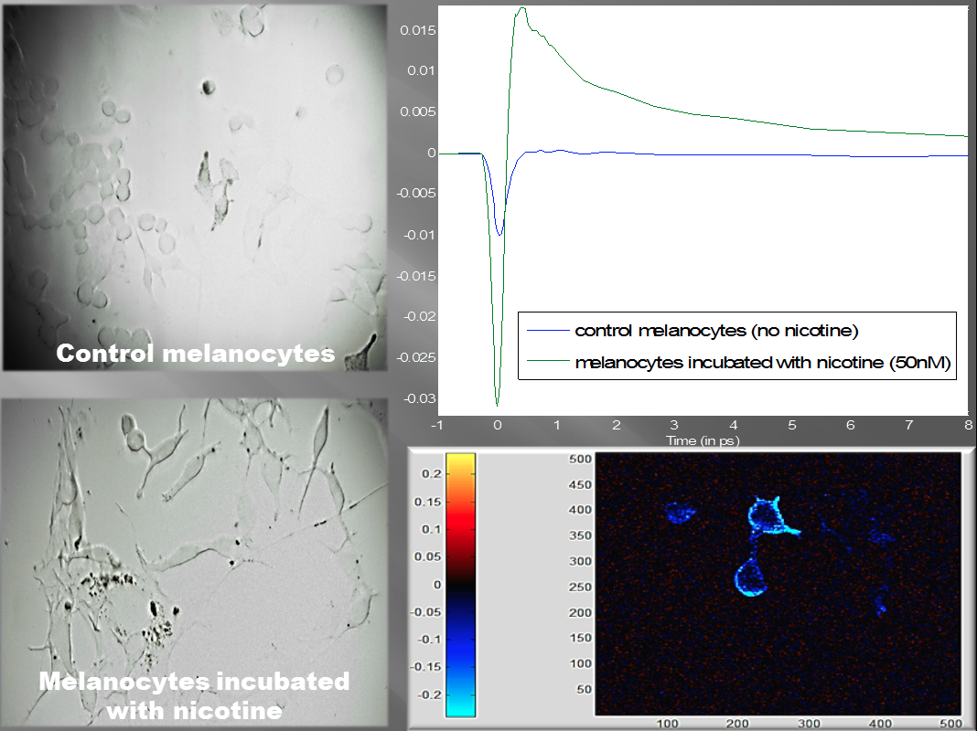
The Center for Molecular and Biomolecular Imaging provided pilot funding to Research Associate Christopher Dall to further explore the relationship between nicotine and melanin in melanoma formation. Preliminary data suggests that nicotine melanocytes exhibit a different structure as well as different optical properties. The melanocytes incubated in nicotine exhibit more and lengthier dendrites (the extensions from the main cellular body). These dendrites can be seen in the bottom left image when compared to normal melanocytes in the top left image.

Melanocytes incubated in nicotine (bottom left) display more dendrites than normal melanocytes (top left). The pump-probe spectra shows more excited-state absorption from the nicotine incubated melanocytes (top right). A pump-robe image shows melanin localizing around aberrant cancerous cells (bottom right).
Pump-probe microscopy may be useful in looking at metabolism of nicotine in humans. In addition, this may give insights into the chemistry, localization, and properties of melanin that are protective against diseases affecting melanin-containing tissues, such as Parkinson’s disease or melanoma. The data collected in this pilot study helped lead to a supplemental award from the Duke Translational Research Institute (Imaging New Molecular Targets for Melanoma Staging).
This project is now supported by the National Center for Advancing Translational Sciences of the National Institutes of Health under Award Number UL1TR001117.
